Column · @edgartmqs284
How Digital Imaging Is Transforming Cosmetic Dentistry
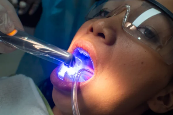

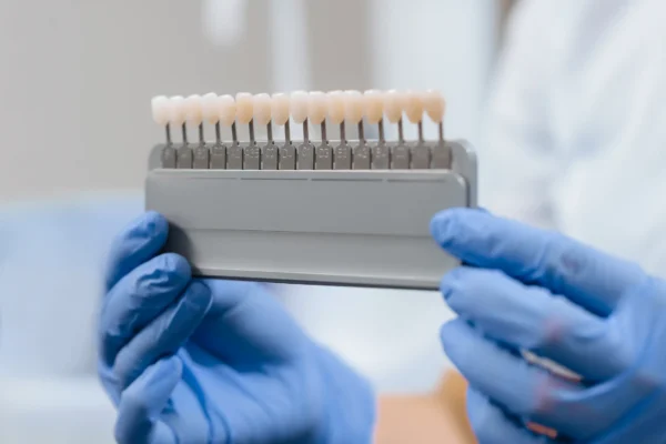
Cosmetic dentistry has always balanced art and biology. Patients come in asking for a brighter smile, straighter edges, less gum display, or a front tooth that does not draw attention the moment they speak. The dentist, meanwhile, has to translate those wishes into something that works with enamel thickness, bite forces, lip movement, facial symmetry, and long-term oral health. For years, much of that translation relied on physical impressions, stone models, shade tabs, and a lot of chairside explanation. Good dentists produced excellent work, but there were limits to how clearly they could show the path from concern to result.
Digital imaging has changed that conversation. It has not replaced clinical judgment, and it certainly has not turned cosmetic dentistry into a simple software exercise. What it has done is make diagnosis sharper, treatment planning more precise, and communication far easier for everyone involved. Patients can often see problems, options, and likely outcomes before a drill ever touches a tooth. Dentists can compare records over time, collaborate more accurately with labs, and catch details that might have been missed on older systems.
In daily practice, the biggest difference is not just speed. It is confidence. When the records are clearer, the plan tends to be clearer. That matters in cosmetic work, where fractions of a millimeter can affect appearance, phonetics, and comfort.
Why images now drive the first conversation
A cosmetic consultation used to depend heavily on verbal description. A patient might say, “My front teeth look short,” or “This crown does not match,” and the dentist would examine, explain, and perhaps sketch possibilities. Today, high-resolution digital photographs and scans bring immediate specificity to that conversation.
A close-up photo of a smile often reveals details patients have noticed for years but never had language for. Incisal wear becomes obvious. A slight midline shift stops being a vague feeling and becomes visible. The way one central incisor reflects light differently from the other can explain why a smile feels “off” even when the teeth are healthy. Once people see their own smile enlarged on a screen, they understand their concerns differently. They stop speaking in broad terms and start responding to concrete features.
That has practical value. Cosmetic dentistry works best when patient expectations are realistic and shared. A person asking for whiter teeth may actually be reacting to uneven edges. Another who thinks they need veneers may improve dramatically with whitening, orthodontic alignment, and bonding. Digital imaging helps uncover the real issue beneath the first request.
There is also a trust element. Patients are more comfortable moving forward when the diagnosis is visible rather than abstract. They can see the crack line, the worn edge, the old composite staining, or the asymmetry in gum height. The conversation becomes collaborative instead of persuasive.
The rise of digital photography as a clinical tool
It is easy to think of smile photography as marketing material, but in experienced hands it is a serious diagnostic instrument. Standardized extraoral and intraoral photographs allow a dentist to study not just teeth, but the context around them. Lip posture, smile line, gingival display, facial proportions, and tooth visibility at rest all influence cosmetic decisions.
A patient may ask for longer central incisors, for example. That request sounds simple until photographs show a high lip line and asymmetric gingival margins. Lengthening the teeth alone might make the smile look bulky. A better plan could involve minor periodontal contouring, selective edge refinement, and restorative changes designed around the full face rather than the teeth in isolation.
Digital photography also helps with shade and texture communication. Natural-looking restorations are not simply one color. They have translucency at the incisal edge, subtle internal character, surface anatomy that reflects light in specific ways, and value that must suit the patient’s complexion and adjacent teeth. A skilled ceramist can do much more when they receive sharp, well-exposed images rather than a written note that says “A1 but slightly brighter.”
In cosmetic cases involving a single front tooth, this can be the difference between a restoration that disappears into the smile and one that looks technically correct but visually obvious. That sort of mismatch used to happen more often than dentists like to admit. Good photography reduces those disappointments.
Intraoral scanners changed more than the impression tray
Among the most visible advances in digital imaging is the intraoral scanner. Patients notice it immediately because it replaces, in many cases, the old putty or polyvinyl impressions that many people dreaded. From the clinician’s side, the impact is even greater.
A modern scanner captures a detailed three-dimensional model of the teeth and surrounding structures. For cosmetic dentistry, that digital model opens up a level of precision and flexibility that traditional impressions could not always deliver consistently. There is no risk of a tray pulling material away from the margin during removal, no bubbles in the impression material, no distortion from delayed pouring, and no shipping damage to a physical model.
That does not mean every scan is automatically perfect. Soft tissue control still matters. Moisture control still matters. A poorly prepared margin is still a poorly prepared margin, whether captured digitally or physically. Yet when the scan is done well, the information can be remarkably accurate.
The practical effects show up across the workflow. Veneers can be planned more conservatively because the dentist and lab can assess contours with confidence. Clear aligner cases tied to cosmetic goals can be integrated into restorative planning more smoothly. Provisional restorations can be designed with better fit. Remakes often decrease because the lab is working from cleaner data.
Patients feel the difference too. Many appreciate being able to watch their teeth appear in real time on a screen. For someone considering veneers or bonding, seeing a 3D map of rotations, spacing, and wear patterns often makes the treatment rationale far easier to grasp than looking in a mirror.
Smile design moved from imagination to simulation
One of the most influential developments in cosmetic dentistry is digital smile design, a broad term that covers software-based planning tools used to analyze facial and dental proportions and create visual treatment simulations. There is a lot of variation in how practices use these tools. Some keep it simple, using photo overlays to discuss tooth shape and symmetry. Others combine facial photography, video, digital scans, and CAD design software to build highly detailed mockups.
Used thoughtfully, digital smile design is not about selling an idealized result. Its value lies in planning and alignment. The software allows the dentist to test proportions before irreversible treatment begins. Width-to-length ratios, incisal edge positions, smile arc, gingival levels, and the relationship between the dental midline and facial landmarks can all be evaluated more deliberately.
This matters especially in veneer cases, where the temptation to chase a trendy look can lead to overbuilt or unnatural restorations. Digital planning can show, early on, whether broader teeth will overpower a narrow face, or whether aggressive lengthening will affect speech. It encourages restraint when restraint is appropriate.
There is a phrase many experienced cosmetic dentists repeat to younger colleagues: just because you can make the teeth larger on a screen does not mean you should. Digital tools are excellent at generating possibilities. Clinical judgment is what keeps those possibilities believable.
A useful simulation can also prevent misunderstandings. If a patient says they want “Hollywood teeth,” that description could mean very different things. One person may want a brighter shade with subtle edge refinement. Another may imagine opaque, extremely uniform veneers. Showing a visual direction early helps uncover those expectations before preparation begins.
Seeing the bite, not just the front view
Cosmetic work is often judged from the front, but it succeeds or fails in function from every angle. Digital imaging has improved the ability to connect esthetics with occlusion, which is where many difficult cases live.
A smile can look beautiful in still photographs while being unstable in motion. If the bite places excessive force on thin ceramic edges, chipping becomes more likely. If anterior guidance is ignored, a patient may end up with restorations that feel foreign or fracture under routine use. If wear patterns are not documented well, the cosmetic result may relapse because the underlying functional problem was never addressed.
Digital scans, occlusal analysis Toothworks of Bakersfield, Dentist and Orthodontist Cosmetic Dentistry systems, and mounted digital models give dentists a better way to study how the teeth meet and move. This becomes particularly important in full smile makeovers, patients with parafunctional habits, or anyone with significant erosion and attrition. The best cosmetic result is not the brightest or most symmetrical one. It is the one that still looks good years later because it was built around a stable bite.
In practice, this often changes the sequence of treatment. A dentist may recommend orthodontic movement before veneers, or protective equilibration, or a night guard after delivery, not because they are adding steps for the sake of complexity, but because the imaging shows the risk clearly. Patients usually accept those recommendations more readily when they can see the contact patterns and wear evidence for themselves.
CBCT and the parts of the smile you cannot see directly
Cone beam computed tomography, or CBCT, is not necessary for every cosmetic case, and it should not be used casually. Radiation exposure, while generally lower than medical CT, still requires justification. But in the right situations, CBCT adds information that flat images cannot provide.
When implant-supported cosmetic dentistry is being considered, CBCT is often indispensable. It reveals bone volume, root positions, anatomical limitations, and angulation possibilities. That information affects not only whether an implant can be placed, but whether the final crown will emerge in a natural-looking position. A front tooth implant can be surgically successful and still cosmetically disappointing if the three-dimensional planning is poor.
CBCT can also help in cases involving impacted teeth, root resorption, complex asymmetry, or prior trauma. A patient may present wanting cosmetic improvement of a discolored front tooth, for instance, but the scan shows structural compromise that changes the prognosis entirely. Better to discover that before a veneer plan is set in motion.
In interdisciplinary cases, CBCT supports cleaner communication between the restorative dentist, oral surgeon, periodontist, and orthodontist. Each specialist can plan from the same anatomy rather than from assumptions. That tends to reduce surprises, and surprises in cosmetic dentistry are rarely welcome.
Lab communication has become far more precise
If you ask dentists where cosmetic cases go wrong, many will eventually point to communication breakdowns. The most elegant preparation and most thoughtful design still depend on the laboratory understanding exactly what the clinician intends. Digital imaging has dramatically improved that handoff.
A modern lab prescription may include high-resolution portraits, retracted intraoral images, stump shades, videos of speech and smile dynamics, digital scans, and annotated reference points. The ceramist can study line angles, texture, embrasures, gingival architecture, and neighboring tooth character in a way that was difficult with traditional records alone.
This is especially important for anterior work. A central incisor is not just a shape. It has personality created by tiny details: the angle of the distal line, the way the light breaks near the incisal third, faint craze lines, the depth of translucency, and the subtle asymmetry that keeps natural teeth from looking machine-made. Better images allow those details to be replicated rather than guessed.
From experience, some of the most successful veneer and crown cases are not the ones with the most advanced software. They are the ones where the dentist gives the lab enough visual information to think like a partner instead of a fabricator. Digital systems make that easier, but only if the clinician uses them well.
Mockups became more persuasive because they became more real
Patients often struggle to commit to cosmetic treatment because they cannot picture the endpoint. Even when they understand the diagnosis, they worry about looking unnatural, too white, too square, or simply unlike themselves. Digital imaging helps bridge that uncertainty through mockups and provisional previews.
A digital wax-up can be turned into a printed model or a matrix that allows a temporary mockup to be placed in the mouth. That means the patient does not just see a simulation on a screen. They can view proposed tooth length, contour, and smile line in the mirror, while speaking and smiling. This step has enormous value.
It also reveals issues early. A design that looks elegant on a monitor may interfere with certain speech sounds or feel too dominant in the face. A patient may realize they prefer slightly softer incisal corners or less overall brightness. These are not setbacks. They are precisely the refinements that make final results feel personal rather than generic.
When digital planning feeds into physical mockups accurately, the treatment process becomes less speculative. That reduces anxiety for the patient and lowers the chance of major dissatisfaction later.
What digital imaging does not solve
It is tempting to talk about technology as if it smooths out every challenge. It does not. Digital imaging improves information, but information still has to be interpreted. Cosmetic dentistry remains vulnerable to overtreatment, trend-driven decision making, and unrealistic expectations.
A scan cannot tell a dentist when a patient would be better served by whitening and orthodontics rather than eight veneers. A smile design simulation cannot guarantee that a patient will emotionally connect with the result. A beautiful set of digital records cannot compensate for weak adhesive technique, poor margin placement, or inadequate understanding of facial esthetics.
There are also practical constraints. Systems cost money, and those costs eventually influence fees. Learning curves are real. Not every practice uses the technology with the same level of skill. A patient hearing “digital smile design” should not assume excellence any more than they would assume excellence from a good camera alone. The operator matters.
Then there is the issue of overpromising. Some simulations are intentionally idealized and can create expectations that biology will not support. Soft tissue may heal differently. Tooth reduction may need to be more conservative than a rendering suggests. Existing restorations, bite constraints, or enamel limitations may narrow the options. The most ethical clinicians frame digital previews as planning tools, not promises.
Where the patient experience has improved most
For patients, the transformation is often less about technology itself and more about how the process feels. Cosmetic dentistry used to require a certain amount of faith. You trusted the dentist’s eye, the lab’s craftsmanship, and the process as a whole. Many people still do, but digital imaging makes trust easier because the process is more transparent.
Several changes stand out in day-to-day care:
- Consultations are easier to understand because visual records replace vague explanations.
- Impressions are often more comfortable when digital scans can be used instead of traditional trays.
- Treatment planning is more collaborative because patients can react to images, mockups, and simulations.
- The lab process is more predictable when detailed digital files support shade and shape communication.
- Long-term comparisons are clearer because records can be reviewed side by side over time. Cosmetic Dentistry
That last point is underrated. In cosmetic dentistry, maintenance matters. Teeth continue to age, restorations continue to face wear, and soft tissue can shift. Having clear digital records from the start helps dentists monitor how a case matures. That can guide polishing, replacement timing, whitening touch-ups, and preventive adjustments before a small issue becomes a visible one.
The conservative side of better imaging
One of the healthiest trends driven by digital imaging is a renewed appreciation for conservative treatment. When the dentist can visualize the case more precisely, there is often less pressure to overprepare or overtreat.
Take edge bonding for worn incisors. In the past, a patient might have been steered toward veneers because the cosmetic endpoint seemed easier to control that way. With excellent imaging, detailed mockups, and careful additive planning, many of those same patients can be treated with far less removal of natural tooth structure. The dentist can preview the esthetic gain and assess the bite before committing to a minimally invasive approach.
The same principle applies to aligning teeth before restoring them. Digital scans and simulations can show that a few months of orthodontic movement may dramatically reduce the amount of enamel reduction needed for cosmetic correction. That is a meaningful shift. Good cosmetic dentistry should not just make teeth look better this season. It should preserve options for the next twenty years.
What this means for the future of cosmetic dentistry
The future is not about replacing dentists with software. It is about integrating better visual data into better clinical decisions. The strongest cosmetic practices are moving toward workflows where photography, scanning, facial analysis, lab collaboration, and provisional testing all support one another. That creates a more complete picture of the patient, literally and biologically.
As these tools continue to improve, the standard for case planning will rise. Patients will expect to see more before treatment begins. Labs will expect better records. Dentists will be able to compare options with greater clarity and defend conservative choices with stronger evidence. The result should be fewer surprises and more durable, individualized outcomes.
Cosmetic dentistry has always demanded an unusual blend of precision and taste. Digital imaging does not remove that demand. It sharpens it. The dentist still needs the eye to judge proportion, the restraint to preserve tooth structure, and the experience to know when a beautiful plan on screen will or will not work in the mouth. What the technology provides is a clearer window into the case, and in this field, clarity changes everything.
Toothworks of Bakersfield, Dentist and Orthodontist
1030 H St #1, Bakersfield, CA 93304, United States
+1 661-323-9421
FAQ About Cosmetic Dentistry
Is bonding better than veneers?
Neither option is best for everyone. Composite bonding can address small chips, gaps, or shape concerns and is generally easier to repair. Porcelain veneers may offer greater resistance to staining and wear, but often require removal of some enamel. A dentist should assess your oral health, bite, goals, and budget before recommending treatment.
How long will dental bonding last?
Bonding commonly lasts about three to ten years before it needs repair or replacement, though this varies with the location and amount of bonding, your bite, and daily habits. Regular dental visits, good oral hygiene, and avoiding chewing hard objects can help protect it. Ask your dentist what lifespan is realistic for your particular treatment.
What are the risks of teeth whitening?
Whitening can cause temporary tooth sensitivity and gum irritation. Overuse or incorrect use may damage teeth or gums, and whitening does not change the color of existing crowns, veneers, or fillings. Have a dentist assess your teeth and gums, and follow the recommended product directions and treatment schedule.